Mole Examination and Mole Removal

Ortalama kalış süresi
Hastanede kalış süresi
Operasyon Süresi
Anestezi Türü
İyileşme süresi
What is a Mole?
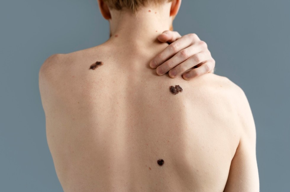
Moles are formations that appear on our body from birth or later, that can be brown, red or skin-colored, that do not cause any symptoms, and that vary in size and thickness depending on the person. Moles have various causes and types. Some moles can be dangerous and cause skin cancer. Therefore, it is important to check your moles regularly and consult your dermatologist if you notice any suspicious changes. Mole removal is a procedure that can be done for both health and aesthetic purposes. The belief that mole removal is harmful is not true. Mole removal should be done with the recommendation and follow-up of the dermatologist. In this article, we will give you information about mole examination and mole removal as YND HEALTH.

Types of Moles
- Melanocytic moles: Moles that originate from cells called melanocytes. They are brown or black in color. Among them, the most common ones are freckle moles, congenital moles and dysplastic moles.
- Vascular moles: Moles that originate from vascular cells. They are red in color. Among them, the most common ones are cherry moles, star moles and port wine stains.
- Epidermal moles: Moles that originate from the cells of the upper layer of the skin. They are skin-colored or light brown. Among them, the most common ones are skin tags, seborrheic keratoses and warts.
Causes of Moles
Genetic and environmental factors are effective in the formation of moles. Some moles are inherited and appear until the age of 30. Some moles develop later and can be seen at any age. Sunlight is the most important environmental factor that triggers the formation and change of moles. Sunlight stimulates the melanocytes in the skin and increases the pigment called melanin. This causes the moles to darken and grow.
The Danger of Moles
- If the color, shape, size or surface of the mole changes
- If the borders of the mole become unclear
- If there is bleeding, crusting, itching or pain on the mole
- If there is redness, swelling or inflammation around the mole
- A: Asymmetry (The two halves of the mole do not match)
- B: Border (The border of the mole is irregular or jagged)
- C: Color (The color of the mole is not even or there are multiple colors)
- D: Diameter (The diameter of the mole is larger than 6 mm)
- E: Evolution (The mole changes over time)
Risk Group for Moles
- Light-skinned, light-haired and light-eyed
- Those who have a family history of skin cancer
- Those who have had sunburns in childhood or adolescence
- Those who have a weak immune system
- Those who are exposed to sunlight or solarium
- Those who have many moles on their body
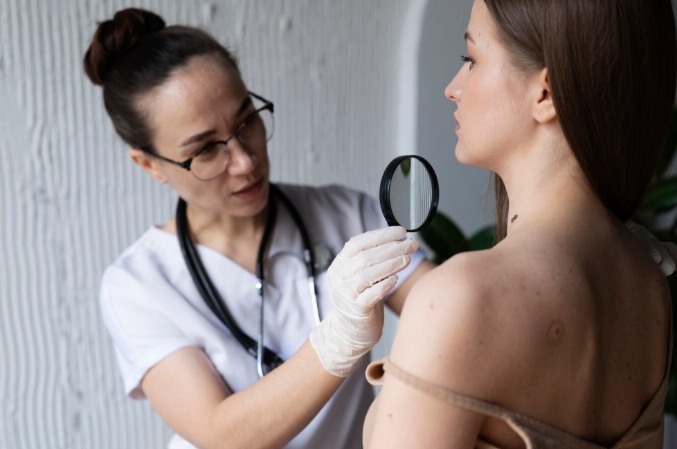
Mole Examination Methods
- Dermoscopy: The method of examining the surface of the mole by magnifying it. It shows the color, shape, border and structure features of the mole. It is done with a device called a dermatoscope.
- Mole mapping: The method of photographing and recording the whole or specific areas of the body. It allows to track the number, location and change of moles. It is done with a device called a digital dermatoscope.
- Pathological examination: The method of examining the mole under a microscope after removing it. It shows the cellular structure and cancer signs of the mole. It is done with a procedure called a biopsy.
Mole Removal Procedure
- Avoiding or protecting from the sun by using high-factor sun creams, hats, glasses and umbrellas
- Checking your moles yourself once a month, observing every part of your body with the help of a mirror, noting the changes in your moles
- Going to the dermatologist once a year for those in the risk group, having dermoscopic examination or mole mapping
- Consulting your dermatologist as soon as you see a suspicious mole, having mole examination and mole removal procedures
- Following your dermatologist’s recommendations on wound care and scarring risk after mole removal
