Eye Diseases
Myopia
Myopia is a condition of blurry distance vision. It occurs when light rays entering the eye focus in front of the retina. It is often a hereditary trait.
Hyperopia
Hypermetropia, or hyperopia, is a condition where near vision is blurred. It is primarily caused by structural and hereditary factors.
Astigmatism
Astigmatism is an eye condition that causes blurry vision at all distances. Degenerative diseases, inflammation, and injuries that cause a deterioration in the structure of the cornea can also lead to astigmatism.
Cataract
Cataracts occur when the lens inside the eye becomes cloudy and no longer transparent. A non-transparent lens prevents light from entering the retina, leading to vision loss.
Cataracts usually occur in the following cases:
✔ In diabetic patients
✔ In congenital cases
✔ In cases resulting from eye trauma
✔ In individuals using corticosteroid medications for an extended period
Cataracts can only be treated through surgery. In the phacoemulsification method, a very small incision is made in the eye, and the cloudy lens is cleaned out. Then, the patient’s natural lens is replaced with an artificial lens. Thanks to phacoemulsification, the surgery is relatively quick and does not require stitches, which accelerates post-operative recovery.
Glaucoma (Eye Pressure)
Glaucoma occurs when the intraocular pressure rises, leading to damage to the optic nerve and loss of the visual field.
In this condition, a person’s visual field gradually becomes smaller. If left unnoticed and untreated, it can lead to blindness.
Before starting treatment, many factors such as the level of intraocular pressure and the extent of optic nerve damage are taken into consideration.
Treatment options can include medications, laser therapy, or surgery.

Retinal Diseases
The retina of the eye is the innermost layer that covers the inside wall of the eyeball and contains the visual cells; it is the deepest layer.
Visual cells are present on the retina. 95% of vision occurs in the macula, which is located in the center of the retina.
Diseases that occur in the retina directly affect vision. The most common symptoms of retinal diseases are:
Sudden or gradual loss of vision.
Distorted or deformed vision.
Flashes of light.
Black spots floating in front of the eyes.
Blurred vision.
Temporary and short-term vision loss.
Retinal Diseases
The retina is the network layer that covers the posterior wall of the eyeball like a wallpaper and consists of visual cells, which is the innermost layer.
It has visual cells on it. 95% of vision occurs in the macula lutea in the center of the retina.
Diseases that occur in the retina directly threaten our sense of sight.
The most common symptoms of retinal diseases are:
✔Sudden or slow vision loss
✔Broken or crooked vision
✔Flashes of light
✔Dark objects floating in front of the eye
✔Screening of the view
✔Transient and short-term vision loss
Retinal Hemorrhages
Inside the eye, there is a clear gel-like fluid called the vitreous body. Blood may leak into this fluid due to various diseases (diabetes, high blood pressure, psychological trauma, etc.). In this case, the vitreous body becomes filled with blood, weakening the patient’s vision and making it difficult for the doctor to detect the retinal problem causing the bleeding inside the eye.
Patients suffering from vitreous hemorrhage often experience sudden vision loss. If the bleeding is mild, patients may see images such as flying objects or rain of dots in their eyes. Causes of retinal hemorrhage include diabetes, high blood pressure, and tears or holes in the retina.
If there is no shock or retinal detachment, no treatment is applied for some time as there is a possibility that the vitreous hemorrhage may heal on its own. During this process, patients are advised to drink plenty of water and keep their heads elevated.
However, if the bleeding does not subside, surgical intervention may be needed to treat vitreous hemorrhage. This surgery, which provides treatment by cleaning the vitreous body, is called Pars Plana Vitrectomy.
Retinal Detachment and Tears
Retinal tear most commonly occurs in high myopia, as a result of thinning of the retinal layer due to the enlargement of the eyeball. However, retinal tears may occur in eyes without defects after trauma or heavy lifting.
Retinal thinning may occur after previous retinal infections and retinal tears may develop from these regions.
The retina is in contact with the vitreous gel that covers the inside of the eye. In a healthy eye, the vitreous is homogeneous, gel-like, and supports the retina. As the vitreous ages over time, the consistency of this gel structure changes and becomes liquid.
The liquefied intraocular gel loses its retina-supporting properties and separates from the retinal layer to which it is attached.
This separation is called posterior vitreous detachment. This disintegrating vitreous gel ripples in the eye with eye movements and is expressed by patients as floating black spots or tulles.
As the intraocular gel separates from the retina, it creates shrinkage. The mechanical stimulation that occurs in the retina as a result of these shrinkages is perceived by the patient as flashes of light or lightning.
These shrinkages can cause tears in the thinned retina. If the torn retinal layer passes over a vessel, there may be bleeding into the eye.
Retinal detachment occurs when the liquefied intraocular gel enters the tear formed in the retina and separates the nerve layer of the retina from the layer to which it is attached. The visual function of the retinal region, which is separated from the underlying tissue, decreases.
Retinal tears that have not developed retinal detachment are surrounded by an argon laser to prevent
Fluid leakage is prevented by surrounding retinal tears which have not developed retinal detachment, with an argon laser. In this case, the vitreous gel is cleaned with the vitrectomy technique, and the inside of the eye is filled with special buffers.
Retinal Vascular Occlusions
It manifests itself with sudden, painless, and persistent vision loss. Depending on the vein that the disease is involved in, there may be a minimal decrease in vision, as well as very serious vision loss.
The risk factors that play a role in the formation of retinal vascular occlusion are:
✔Aging
✔Smoking
✔High blood pressure and diabetes
✔Glaucoma (increased intraocular pressure)
If a part of the retinal vein is blocked and there is fluid accumulation in the visual center, laser and intraocular cortisone treatment is applied.
Macular Degeneration
Macular degeneration is a condition where the macula, a part of the retina, becomes damaged with age, leading to weakened vision.
Patients may see straight lines as wavy. They cannot see clearly where they are looking, but they can see their surroundings. Reading and near vision are the most affected areas.
There are two types of the disease: dry and wet.
In dry macular degeneration, blood vessels are intact, and there is no bleeding. Vision loss occurs, but complete blindness does not develop.
In wet macular degeneration, blood vessels are weak, and bleeding occurs. There is a risk of blindness. Argon laser, photodynamic therapy, and intraocular injection methods are used in the treatment of wet macular degeneration.
Macular Degeneration Treatment
Dry-type treatment is done with oral support drugs. In wet-type treatment, argon laser, photodynamic therapy, and intraocular injections are used. In photodynamic therapy, the capillaries that bleed in the retina are stained with a special drug administered with a needle inserted into the patient’s arm, and the diseased area is destroyed with a laser applied to the dyed area.
Anti-VEGF drugs are used in intraocular injections. Injection application is easy and painless for the patient. Intraocular injections are made 3 times with an interval of one month.
Patients with reduced vision can also be greatly helped with special glasses called LVA. Most patients can read newspapers comfortably with these glasses. Patients with macula lutea should be examined every 4 months.
Diabetic Retinopathy
It is the most common cause of blindness due to diabetes. Diabetes disrupts the nerve layer of the eye (the retina or the retina) and the blood vessels in this layer, preventing them from performing their function and causing vision loss.
The progression of retinal disorder due to diabetes is called diabetic retinopathy.
Corneal Diseases
The cornea is the transparent curved layer of the eye located in the front part of the eye. It has the function of refracting light and protecting the eye from external factors, and it plays a crucial role in vision, serving as the eye’s most important refractive lens. The cornea can lose its transparency due to various diseases, which may be congenital, hereditary, or microbial.

Keratitis
Corneal inflammation is called keratitis. Keratitis can be caused by microorganisms, viruses, fungi, or rheumatism. Symptoms of keratitis include pain, burning sensation, redness in the eye, blurred vision, and white spots in the eye. Medication is applied as treatment, and in advanced cases, corneal transplantation may be required.
Keratoconus
Keratoconus is a disease that occurs when the cornea becomes gradually thinner and steeper; in other words, it is a deformation of the cornea. Patients are treated by wearing contact lenses or by using corneal ring treatments if they are not suitable for contact lenses, and if the disease is not in an advanced stage. In more advanced stages of keratoconus, the last resort is corneal transplantation.
Dry Eye Syndrome
Dry eye syndrome occurs due to insufficient tears. Symptoms include a stinging sensation, redness, and a constant feeling of a foreign body in the eye. Treatment includes the use of oral supplements for dry type and the use of artificial tears or medications for other types.
Eye Tumors
In cases of white spots in the eye, changes in the color of the iris, and unexplained vision loss, the possibility of an eye tumor should be examined. A family history is important for eye tumors.

Retinoblastoma
Retinoblastoma is a tumor that occurs during infancy and childhood. It is usually noticed through whitening of the eye in one or both eyes. Cross-eye in children is another symptom. Chemotherapy, surgical removal (enucleation), and laser radiation therapy are used in treatment.
Uveal Melanoma
These are tumors that generally appear between the ages of 55-60. Symptoms may include a spot on the iris or a change in the color of the iris in one eye. Those located in the back area may cause complaints such as blurred vision and floaters. In some cases, they can be noticed incidentally during routine eye examinations. Treatment may include surgery and radiation therapy for plaques.
Metastatic Tumors of the Eye
These are tumors that occur in the eye due to the spread of cancer cells from elsewhere in the body. They often cause symptoms such as vision loss. The most common cancers that spread to the eye are breast cancer in women and lung cancer in men.
Eye Aesthetics
Eyelid drooping
Ptosis, a condition in which the upper eyelid is lower than normal, is called a drooping eyelid. This condition can occur congenitally or due to reasons such as aging or eye surgery.
– The causes of ptosis include:
Damage to the nerve that lifts the eyelid.
Weakness or underdevelopment of the eyelid muscle (this muscle is insufficient in congenital ptosis).
Sagging of the eyelid skin due to aging.
Previous eye surgeries.
The treatment for ptosis is surgery.
Under-eye bags
The major reason for bags under the eyes is genetic factors. Under-eye bags can make a person appear older and more tired. In treatment, excess fat tissues are removed or replaced.
Congenital Small or Absence of the Eye
The size and shape of the eye affect the development of the tissues around the eye. If the eye does not grow fully, the eyelids, loose tissues, and bone structure around the eye remain smaller than normal. This condition is surgically corrected.
Diagnostic Methods Used for Eye Diseases

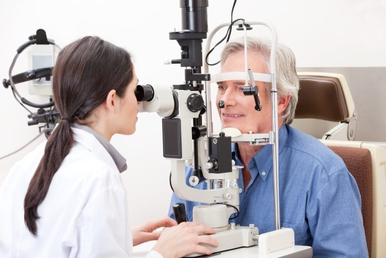
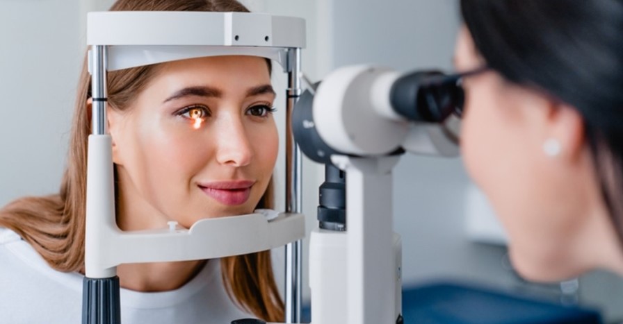
The primary methods used to diagnose eye diseases are:
✔Eye Ultrasonography
✔Pachymetry
✔Wave-front
✔Ultrasonic Biometry
✔Humphrey Visual Field Test
✔Auto Refractometer
✔OCT (Optic Coherence Tomography)
✔Eye Angio (FFA and ICG)
✔Corneal Topography
Eye Ultrasonography
Ultrasound Imaging of the Eye: In eye examinations, ultrasound waves are used to image the part behind the eye. Using a method called B-Scan, conditions such as bleeding inside the eye, foreign bodies, tumors, or retinal detachment can be detected.
Pachymetry
Corneal Thickness Measurement: A device called pachymetry is used to measure the thickness of the cornea, the outer layer of the eye. This device plays an important role before Excimer Laser surgery and in diagnosing glaucoma.
Wave-Front
Wavefront Analysis: Thanks to wavefront technology, a three-dimensional map of the eye is obtained. This map reveals all the defects present in the cornea, allowing for custom laser treatment.
Ultrasonic Biometry
Biometric Measurements Using Ultrasound: In cataract surgery, the damaged part of the lens is removed and replaced with an artificial lens that matches the patient’s eye number. The biometric ultrasound measurement method is used to determine the number of lenses to be placed in the eye.
Humphrey Visual Field Test
Humphrey Visual Field Test: A device called Humphrey is used to perform the visual field test. This device not only measures the area that the eye is looking at but also measures the area located at the edges. This device is very important in the early diagnosis and monitoring of glaucoma.
Auto Refractometer
Auto Refractometry: This device measures the cornea’s ability to refract light. It bends incoming light rays onto the retina. This bending is called refraction.
Eye Angio (FFA and ICG)
Fundus Angiography (FFA and ICG): FFA is a method that reveals the structure of the retina and blood vessels. The central vision and vascular structure of the retina are examined using this method. In this method, a special dye is injected into the patient’s vein.
OCT (Optical Coherence Tomography)
Optical Coherence Tomography (OCT): With OCT, the retina can be imaged in thin slices, allowing for the diagnosis and monitoring of glaucoma in particular.
Corneal Topography
Corneal Topography: Thanks to the Pentacam device, even the finest details can be examined. This device also provides detailed information about corneal thickness. This way, doctors can determine whether the patient’s eye is suitable for laser treatment or if there is any corneal disease.
Treatment Methods Used in Eye Diseases
The main treatment methods used in the treatment of eye diseases are:
✔Corneal Transplantation
✔Excimer Laser
✔Enucleation
✔Evisceration
✔Exenteration
✔Eye Prostheses
Corneal Transplantation
The cornea is the curved, transparent layer of the eye located on the outer part of the eye. It refracts and focuses light and protects the eye. It plays a crucial role in vision as it is the eye’s primary lens. Transparency of the cornea can be lost due to various diseases.
These diseases can be congenital, hereditary, or infectious. The process of replacing damaged corneal tissues with healthy corneal tissues is called corneal transplantation. This procedure is performed under general anesthesia.
Corneal transplantation can be performed using laser technology in addition to the traditional method. With this method, incisions of the required size and depth can be created.

Excimer Laser
The Excimer Laser technique is the most commonly used method in the world for treating eye defects such as myopia, hyperopia, and astigmatism. Laser is also used to remove defects on the surface of the cornea due to disease or injury, scars on the transparent layer, corneal transplantation, and eye defects that occur after transplantation. Excimer Laser is a very fast, reliable, and completely painless method.
People who are eligible for laser surgery should have the following features:
✔ They are over 18 years old and wear glasses or contact lenses.
✔ Their eye prescription hasn’t changed more than +/- 0.5 diopters in the past year.
✔ People with myopia up to -10 diopters.
✔ People with astigmatism up to -6 diopters.
✔ People with hyperopia up to +5 diopters.
✔ Those with sufficient corneal thickness and a normal corneal structure.
✔ They do not have systemic diseases like diabetes or rheumatism.
✔ There are no other diseases in their eyes.
✔ Not pregnant.
Laser procedures take 5 to 10 minutes, and the patient can resume normal activities after one day. There is no need to cover the eyes with a bandage after treatment. It is important not to touch or wash the treated eye during the first 24 hours.
Laser Technologies
Lasik
LASIK: In this method, a thin flap called a “flap” is created on the outermost surface of the cornea layer, and eye defects are corrected by applying the Excimer laser to the corneal tissue under this flap. This procedure is performed under local anesthesia without the need for needles or sutures. The patient does not feel any pain during or after the operation.
iLasik
iLASIK: iLASIK is a personalized method. First, a vision profile of the person is created using the Wavescan technique. The Wavescan system creates a three-dimensional map of your eye defects. Then, a personalized treatment is planned using the digital data obtained from this map. The corneal flap is then created using IntraLase without a blade. Information reflected from the eye is detected by a special camera. This information is transferred via laser, and the planned treatment is performed.
PTK (Treatment of corneal spots/irregularities)
PTK (Phototherapeutic Keratectomy): Some eye diseases or injuries can cause spots on the cornea. In this case, laser is applied to the eye using a technique called PTK to eliminate these spots. For PTK to be performed, the spots must be on the surface. If the spots are deep in the cornea, PTK cannot be performed, and corneal transplantation is required.
PRK
PRK (Photorefractive Keratectomy): PRK is a method used to correct vision defects. In this method, the superficial tissue, the outer layer of the cornea, is removed, and the shape of the cornea is changed using laser beams.

Enucleation
Enucleation is a surgical technique used to treat malignant eye tumors that arise from places like the eyelid, eye, or inside the orbit and spread to surrounding tissues.
Evisceration
Evisceration is one of the techniques used in cases that require the removal of the eye. In this technique, the hard, white outer layer of the eye is left intact, and unhealthy tissues inside the eye are removed.
Exenteration
Excision is the complete removal of the eye in cases where the eye has lost its function due to various reasons. During the surgery, the muscles that move the eye are preserved, and a spherical implant is placed instead of the eye.
Eye Prostheses
Artificial eyes are devices that resemble the other eye and are used in cases of congenital eye defects or extremely small eyes, following procedures such as excision or evisceration, to ensure facial symmetry. They are typically made of glass or acrylic (a type of plastic).
