Brain and Nerve Surgeries
It is a branch of surgery that performs operations to treat problems in the nervous system. Doctors working in this field should have extensive experience because the nervous system is highly sensitive and complex.
Main areas of interest in neurosurgery:
✔ Brain tumors
✔ Spinal tumors and myeloma
✔ Aneurysm
✔ Narrowing in neck veins
✔ Herniated disc
✔ Cerebral hemorrhage
✔Epilepsy
✔Parkinson’s disease
✔Scoliosis
✔ Nerve compressions
✔ Stroke

Brain Tumors
When cells in the brain grow differently or irregularly than normal, it is referred to as a brain tumor. The following symptoms are the most common in people with brain tumors:
✔ Headaches
✔ Visual disturbances
✔ Seizure-like fainting
✔ Partial paralysis in some parts of the body
✔ Severe vomiting
✔ Personality changes and significant mood swings
✔ Weakness and numbness
✔ Contractions
✔ Difficulty understanding
✔ Balance problems
✔ Loss of coordination
✔ Brain tumors can be benign or malignant. Characteristics of benign brain tumors are:
✔ They can be removed through surgery and usually do not recur.
✔ They do not spread to other parts of the brain.
✔ They can be life-threatening by pressing on the surrounding area.
✔ In rare cases, a benign brain tumor can transform into a malignant brain tumor.
Malignant brain tumors can be defined as follows:
✔ They contain cancerous cells.
✔ They grow rapidly and infiltrate healthy tissues.
✔ Sometimes they can spread to the spinal cord or other parts of the body, meaning they metastasize.
✔ Whether benign or malignant, the most effective treatment method is surgery.
Spinal Cord and Spinal Column Tumors:
Tumors that develop in the bones, nerves, or soft tissues that make up the spinal column and spinal cord are called spinal cord and spinal column tumors. Spinal cord tumors may originate from the spinal column and spinal cord themselves, or they may occur when tumors spread from other parts of the body (such as the breast, prostate, lungs, etc.) to the spinal column.
The exact cause of spinal cord and spinal column tumors is not fully known. Genetic factors, exposure to certain chemicals, radiation exposure, and viral infections are considered risk factors that play a role in the formation of these tumors.
The most common symptoms in people with spinal cord and spinal column tumors include:
✔ Back pain that doesn’t go away with rest
✔ Fever, weight loss, fatigue
✔ Weakness and numbness in the arms and legs
✔ Urinary or bowel incontinence
✔ Sexual dysfunction
✔ Loss of reflexes
✔ Apathy, bedridden state
✔ Impairment, weakness, and seizures in children
Radiological imaging techniques are used to diagnose spinal cord and spinal column tumors. Once a tumor is found, a biopsy is taken to determine its type and source.
Different branches of medicine, such as neurosurgery, neurology, radiation oncology, medical oncology, and pathology, collaborate in the treatment of spinal cord and spinal column tumors. Depending on the location and type of tumor, treatment methods such as surgery, radiation therapy, chemotherapy, and immunotherapy can be applied alone or in combination.
Dilation of blood vessels (Aneurysm)
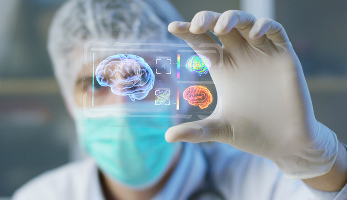
Cerebral blood vessel dilation is a permanent swelling that occurs in the blood vessels of the brain. Factors such as genetics, age, and gender play a role in the development of these swellings. Most cerebral blood vessel dilations are discovered as a result of the tearing of blood vessel dilation and bleeding. This bleeding is very severe and can lead to a cerebral hemorrhage. The mortality rate due to cerebral blood vessel dilation bleeding approaches 50%.
25% of patients who survive cerebral blood vessel dilation bleeding experience permanent nerve damage and body function impairment, such as paralysis. Cerebral blood vessel dilation typically does not show any symptoms before bleeding occurs. When bleeding occurs due to cerebral blood vessel dilation in the brain’s blood vessels, the person experiences severe and continuous headaches they have never had before.
In addition to this pain, there are also symptoms such as nausea, vomiting, neck stiffness, light sensitivity, and numbness.
Today, there are three important treatment methods for patients diagnosed with cerebral blood vessel dilation:
✔ Monitoring and non-surgical treatment (monitoring only): If cerebral blood vessel dilation has not torn in the brain’s blood vessels, the only treatment option is medication. Regulating blood pressure is the primary goal in drug therapy. It is essential to use medications that lower blood pressure and support treatment with a healthy diet and an exercise program.
✔ Surgical treatment and occlusion (clipping) of cerebral blood vessel dilation: This is a permanent treatment method. A small part of the skull is opened, and a metal clip is placed between the vessel containing cerebral blood vessel dilation and the cerebral blood vessel dilation itself, thereby preventing blood from entering the cerebral blood vessel dilation.
✔ Stents and endovascular treatment within blood vessels: Inside the blood vessels, the principle is to use vascular imaging machines, introduce very thin tubes into the cerebral blood vessel dilation, cover the cerebral blood vessel dilation sac with very flexible metal wires called coils, and cut off the blood flow to the cerebral blood vessel dilation. This is the most preferred method in the world for treating cerebral blood vessel dilation.
Narrowing of the carotid artery (carotid artery stenosis):
Carotid artery stenosis is a narrowing or blockage of the carotid artery, the major blood vessel in the neck. The jugular vein is the vessel that carries blood to the brain. If blood supply to the brain is interrupted, the brain cannot get oxygen and nutrients, leading to a stroke.
If blood flow is not restored, brain cells die quickly, resulting in permanent brain damage. Risk factors for carotid artery stenosis include age, blood pressure, smoking, high cholesterol levels, diabetes, and obesity.
Usually, it does not cause any symptoms. If the narrowing in the carotid artery is less than 50%, blood clot prevention and thinning medications are used. If the narrowing is more than 70%, surgery is the most effective treatment method.
Herniated Disc
There are 5 vertebrae in the cervical region of the spine, and between these vertebrae, there is a special tough tissue called the disc.
A herniated disc occurs when these discs protrude and press on the nerves. Causes of a herniated disc include improper movements during daily activities, excess weight, inactivity, and smoking, which leads to disc degeneration.
The most common symptoms of a herniated disc are:
✔ Lower back and leg pain
✔ Difficulty in movement
✔ In addition to pain, tingling and numbness in the legs
✔ Loss of energy
Surgery is not recommended when a herniated disc first begins. Muscle relaxants are given, and certain movements are advised to be avoided, with frequent rest. If the herniation progresses, physical therapy under the supervision of a specialist doctor is appropriate.
If conservative treatment does not yield results, surgery is necessary. Microscopic surgery is the widely preferred method today. In this method, procedures are performed using an operating microscope. Patients typically stay in the hospital for one day. The risk of recurrence and complications is very low.
Brain Bleed
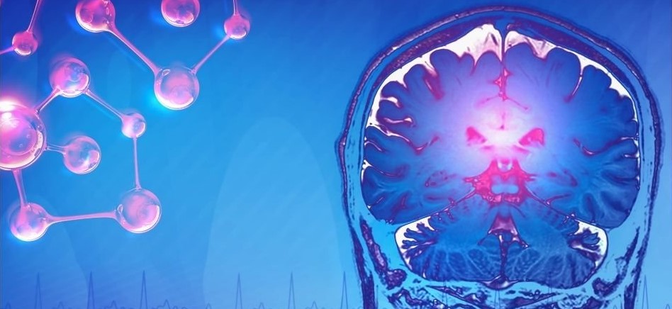
Brain bleed, also known as cerebral hemorrhage, is the leakage of blood into the brain as a result of the rupture of a blood vessel in the brain. There are two types of brain bleeds: bleeding within the brain tissue (intracerebral) and bleeding between the layers (subarachnoid). The most common cause of bleeding within brain tissue is high blood pressure. Bleeding between the brain’s membranes usually occurs when a blood vessel bubble (aneurysm) in the brain bursts.
The most common symptom of a brain bleed is the sudden onset of severe headaches. Nausea and vomiting may also accompany the headache. Gradual loss of consciousness may occur, leading the patient into a coma. There may also be a deterioration in vision, slurred speech, the appearance of blind spots in the visual field, or sudden loss of vision in one eye.
The best way to prevent brain bleeding is to maintain blood pressure at normal levels.
Epilepsy
Epilepsy is a disorder that occurs in the form of seizures due to temporary abnormal electrical activity in the brain cells. The patient may experience loss of consciousness for a certain period of time, body convulsions, fainting, urinary incontinence, hand and arm movements, and more. Changes in behavior, emotions, movement, or perception can also be observed. This condition requires long-term treatment and significantly affects the quality of life. If epilepsy cannot be controlled with medications or if medical treatment is insufficient, surgical approaches can be considered.
There are two main types of epilepsy surgery. The first is the removal of the seizure-causing area (resective surgery). The second is the cutting of pathways of seizures to prevent or reduce the spread of seizures (functional, palliative surgery).
Parkinson’s disease
Parkinson’s disease is a condition that affects older adults, with an increased incidence after the age of sixty. However, the exact cause is not known. Parkinson’s disease leads to a weakening of movement and involuntary movements.
Symptoms include tremors in the hands and feet, slow movements, muscle stiffness, and difficulty walking. The first choice for treating Parkinson’s disease is medication. If medications cannot control the disease or if medical treatment is unsuccessful, surgery may be considered.
There are two surgical approaches used in the treatment of Parkinson’s disease. If the symptoms of the disease are on one side, a technique like laser therapy is applied. If the symptoms affect both sides, a deep brain stimulation device is implanted to regulate brain impulses.
Scoliosis
Scoliosis is the curvature of the spine to the right or left. Usually, scoliosis does not cause symptoms initially, but as it progresses, the following symptoms may appear:
✔ One shoulder is higher than the other.
✔ When the arms hang down on both sides, there is more space between the arm and the body on one side.
✔ One hip appears higher or more prominent than the other.
There is some evidence that scoliosis can be hereditary, but despite all the research, it is not entirely understood what causes scoliosis.
Treatment for scoliosis varies depending on the type and size of the curvature, but the most commonly used treatment methods are:
✔ Observation: In patients whose temperature is below 20 degrees and whose bone structure growth is nearly complete, observation and regular monitoring are usually sufficient.
✔ Bracing: Bracing is used to prevent the progression of scoliosis in children whose curvature is over 25 degrees and who are still growing.
✔ Surgical treatment: When the curvature exceeds 50 degrees and the child is still growing, surgical intervention may be needed to straighten the spine.
Nerve Compressions
It is a common neurological problem.
Factors that can cause nerve compression include:
✔ Injuries and fractures
✔ Cysts and tumors around nerves
✔ Congenital deformities
✔ Rheumatic diseases, thyroid diseases, diabetes, other neurological diseases, blood disorders, gout
✔ Factors such as alcohol addiction, smoking, and obesity
✔ Hormonal changes such as pregnancy and menopause
✔ Occupational diseases (repeated and strenuous joint movements)
✔ Burns
The most common type of nerve compression is “carpal tunnel syndrome,” which affects the wrist and fingers.
In this condition, the nerve extending to the first four fingers of the hand is compressed at the wrist level. This condition is more common in women and in professions that use their hands frequently.
The main symptom of the condition is numbness in the hand. There may also be pain. Patients often shake their hands to relieve numbness. Over time, finger strength decreases, and objects in the hand may be dropped.
A similar problem occurs in the ankles and toes, especially in those who prefer tight-toed shoes and high heels, and those who engage in strenuous activities, known as “tarsal tunnel syndrome.”
A sensitive method called EMG is used to determine the level of nerve compression.
Physical therapy is applied in cases that have not progressed yet and do not involve muscle strength loss.
In advanced cases, surgical intervention is performed to relieve pressure on the nerve.
Nerve Compressions
Trigeminal Neuralgia
Trigeminal neuralgia, also known as tic douloureux, is defined as an extremely severe pain and is very rare in people under the age of thirty.
Trigeminal neuralgia pain occurs due to disorders in the structure of the blood vessels surrounding the trigeminal nerve, which is one of the twelve pairs of nerves that emerge directly from the brain.
Medications are the first choice in treating trigeminal neuralgia pain. If medications are unable to sufficiently relieve the pain or lose their effectiveness, glycerin injections or the burning of painful fibers to disconnect the nerve may be performed. In surgical treatment, the blood vessel compressing the nerve in the brainstem is removed.
Spasms
In the case of spasms, muscles contract continuously. Muscles that are stiff or tight due to contraction interfere with normal functions such as walking, movement, and speech.
Symptoms of spasms include an inability to bend the arms or legs, joints staying at unusual angles, crossing of the arms or legs, and difficulty speaking. If spasms persist for a long period, muscles can press on the joints and force them into a fixed position.
Physical therapy is an important method for treating spasms. Medication can be administered orally or by injection into the affected area. In cases of severe and chronic spasms, surgical treatment may be necessary.
Facial Spasms
Facial spasms are intermittent, involuntary, and painless contractions of facial muscles. They usually start around the eyes and then spread to other facial muscles around the mouth. They can last for seconds or a few minutes.
Medications and Botox are frequently used in mild cases of facial spasms. If Botox treatment fails or if the conditions are advanced and clinical, surgery may be applied.
Stroke
When the blood supply to the brain tissues is interrupted for any reason, often due to blockages in blood vessels in the brain or neck, the brain cells cannot get oxygen and nutrients. If this condition is not resolved quickly, brain cells die, leading to irreversible brain damage.
There are two types of strokes: ischemic stroke and hemorrhagic stroke. Ischemic strokes occur when the blood vessel carrying blood to the brain is blocked. Hemorrhagic strokes happen when blood vessels in or around the brain rupture.
Although strokes are more common in older adults, they can occur at any age.
Key symptoms of a stroke include:
✔ Severe headache
✔ Confusion and altered consciousness regarding person, place, and time
✔ Numbness or weakness in the arm, leg, or face
✔ Sudden slurred speech
✔ Loss of vision
✔ Balance disturbance or an inability to perform tasks requiring coordination
There are various treatment approaches for stroke, which may involve repairing ruptured blood vessels in the brain, removing blood clots forming in the brain, or cleaning plaques in the neck’s carotid arteries to prevent brain blockages.
Diagnostic methods used in neurosurgery
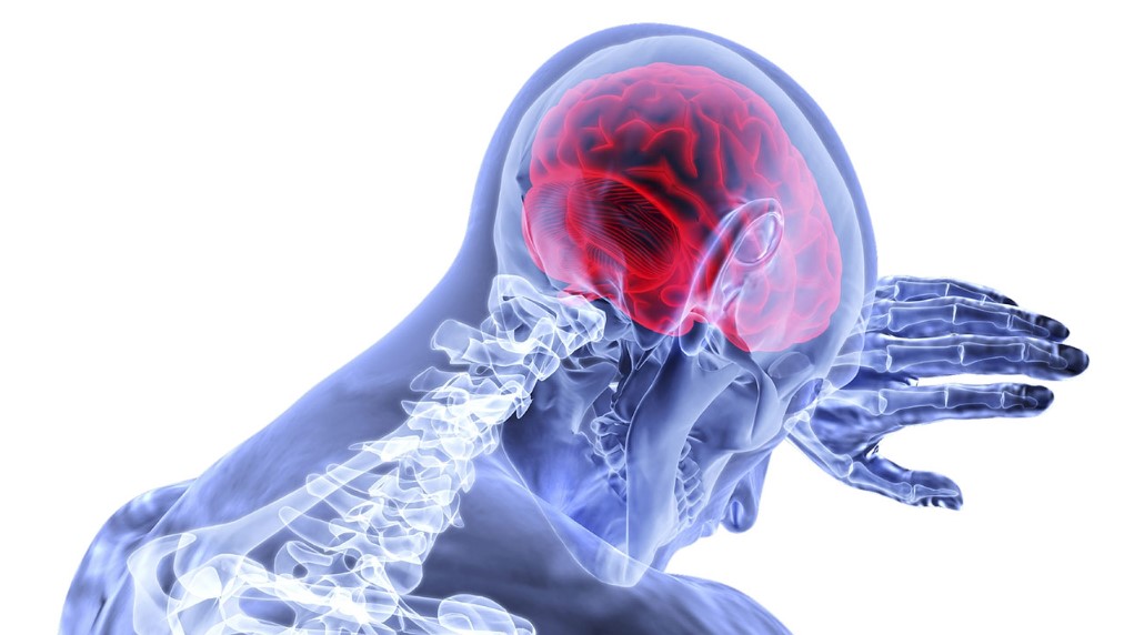
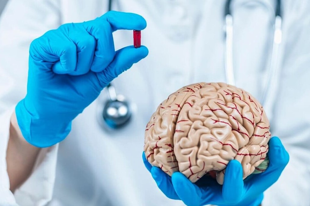
Diagnostic methods frequently used in neurosurgery are as follows:
✔ CT (Computed Tomography)
✔MR Imaging
✔MR Angiography (MRA)
✔Catheter Angiography
✔ Nuclear medicine
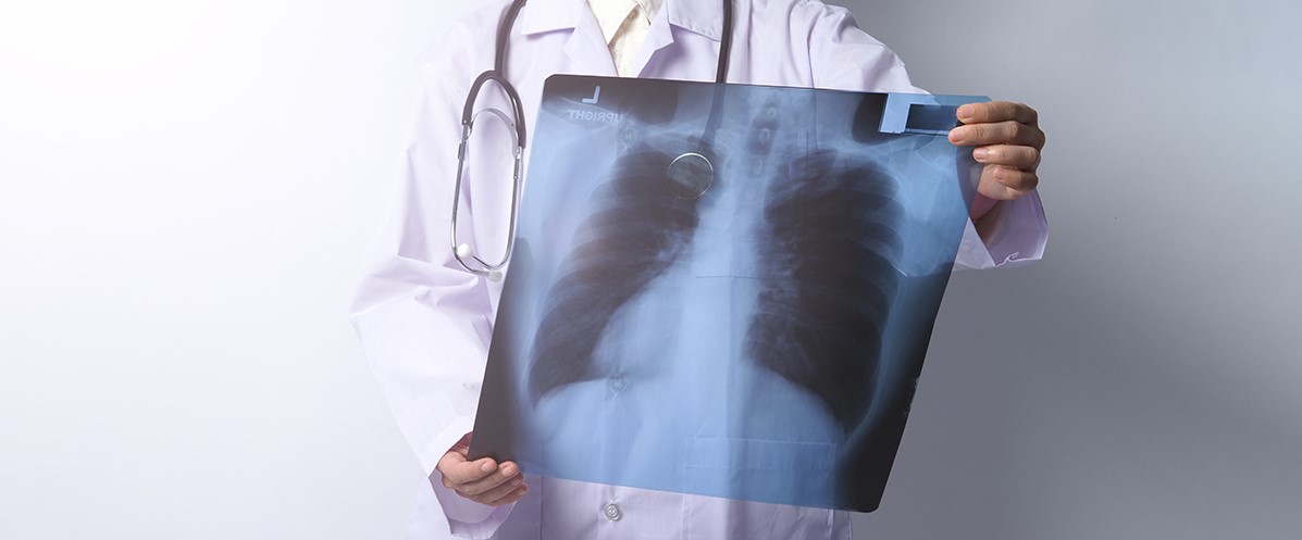
CT (Computed Tomography)
Computed tomography (CT) scanning is a specialized imaging method that uses X-rays to create detailed cross-sectional images of tissues and organs in the body. CT scans can also be used to image bones, soft tissues, and blood vessels.
MR Imaging
Magnetic resonance imaging (MRI) is a non-invasive method that provides highly detailed and clear images of the body’s organs and tissues. Unlike X-rays, MRI does not use ionizing radiation but instead relies on radio waves and a strong magnetic field.
MRI is considered one of the most sensitive techniques for diagnosing brain tumors, strokes, and some chronic neurological disorders (such as multiple sclerosis). It can also be used to image small structures in the eye or inner ear.
MR Angiography (MRA)
Magnetic Resonance Angiography (MRA) is an application of magnetic resonance imaging that displays the blood vessels in the body. MRA is an extremely useful examination for identifying vascular problems and determining the most appropriate treatment method.
With this method, the major blood vessels can be easily visualized without the need for contrast agents. However, by using it in combination with special contrast agents, the fine details of the veins can be enhanced to improve image quality.
Catheter Angiography
Angiography involves the insertion of a catheter into the artery of the area to be examined, followed by the administration of a contrast material to enable X-ray imaging of the blood vessels.
Nuclear Medicine
In nuclear medicine, radioactive substances are used for both diagnostic and therapeutic purposes. In conventional radiology, the source of radiation is external, with the patient positioned between the radiation source and the film.
In nuclear medicine, radioactive materials are administered to the patient orally or through other means to capture images, and the emitted radiation from the patient is detected.
Positron Emission Tomography (PET imaging)
PET imaging is an advanced nuclear medicine technique. With positron emission tomography (PET) imaging, early diagnosis can be achieved by identifying tumor tissues that may not be visible using conventional radiographic methods.
It’s easy to determine whether a tumor is benign or malignant with PET imaging. The stage of the tumor can also be easily determined through PET imaging.
Assessing the tumor’s response to treatment is possible by comparing PET scans before and after treatment. Following radiation therapy, chemotherapy, and surgical interventions, PET imaging can differentiate whether the tumor tissue is alive or not. Additionally, in cases of disease recurrence, the location of recurrence can be identified through PET imaging.
Treatment Methods Used in Brain and Nerve Surgery
Common Treatment Methods in Neurosurgery include the following:
✔ Micro Surgery
✔ Gamma Knife
✔ Endovascular Surgery
Micro Surgery
Microsurgery refers to surgical procedures performed using a microscope. Thanks to the microscope, tissues can be seen more clearly and in greater detail during surgery, making it possible to perform surgery through much smaller incisions. This increases the success rate of surgery by causing minimal damage to healthy tissues and allowing full access to the tissues with problems. Today, most neurosurgery procedures are performed using microsurgical techniques.
Gamma Knife
Gamma Knife is an unconventional method that allows neurosurgeons to perform procedures on abnormal parts of the brain without making an incision. Gamma Knife is a therapeutic system that defines the coordinates of the patient’s brain tissues and destroys them with gamma rays. In this surgical method, the treatment is completed in one session. It is generally used in cases where open brain surgery is risky or impossible.
Endovascular Surgery
Endovascular treatment is applied in cases where the patient may be at risk, depending on the location of vascular dilation. In this method, a thin tube called a microcatheter is inserted from the groin area to access the site of vascular dilation in the brain. Then, a vascular dilatation sack is completely isolated from blood flow by filling it with very thin wires called “coils.” This eliminates the risk of bleeding because blood cannot enter the dilated blood vessels.
