Cardiology
Primary Areas of Concern in Cardiology
✔ Arrhythmias (Irregular Heartbeat)
✔ Heart Attack
✔ Myocarditis (Inflammation of the Heart Muscle)
✔ Pericarditis (Inflammation of the Outer Membrane of the Heart)
✔ Coronary artery disease
✔ Heart Valve Diseases
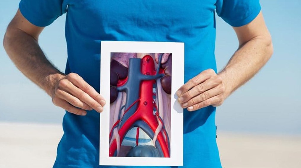
✔ Aortic Aneurysm
✔ Congenital Heart Defects in Children
Arrhythmias (Irregular Heartbeat)
Arrhythmias, or irregular heartbeats, are a condition in which the normal rhythm of the heart is disrupted.
In individuals with arrhythmias, the heart rate may become faster or slower than necessary, and the heartbeats may not be regular.
Causes of arrhythmias include genetic factors, smoking, stress, and caffeine consumption. Sometimes, arrhythmias can be due to congenital heart defects or be associated with later-developing diseases.
Common underlying conditions include high blood pressure, coronary artery disease, and age-related conduction system degeneration.
The most common complaint for patients with arrhythmias is palpitations. Additionally, there is a risk of fainting, dizziness, fatigue, and sudden death.
When medications are not sufficient in treating arrhythmias, a procedure called ablation is performed. In the ablation process, radiofrequency energy is used to heat the affected area and eliminate the source of the irregular heartbeats or palpitations. This method is known as radiofrequency catheter ablation.
Another method is known as cryoablation, where the targeted area is frozen, disrupting its function using nitrogen gas. Both methods have their pros and cons. Therefore, the choice of treatment should be based on the type and severity of the arrhythmia.
Heart attack
A heart attack is a condition in which a part of the heart muscle is damaged due to a disruption in blood flow. Five cases of sudden death are often caused by heart attacks.
Risk factors for experiencing a heart attack include high blood pressure, high cholesterol levels, diabetes, smoking, and a family history of coronary heart disease. Many of these factors are associated with excess weight. The formation of a blood clot in a narrowed artery can occur for various reasons, and sometimes stress can also be a trigger.
The most prominent symptom of a heart attack is chest pain. In addition to chest pain, the following symptoms may also occur:
✔ Chest tightness or burning, which may be mistaken for stomach discomfort. It is often accompanied by cold sweats and fear.
✔ Difficulty breathing
✔ Coughing
✔ Dizziness and lightheadedness
✔ Fainting
✔ Nausea and vomiting
A heart attack is a life-threatening condition. The patient should be urgently admitted to the hospital and transferred to the intensive care unit. The treatment goals are to halt the progression of the heart attack and reduce heart damage.

Myocarditis (Heart Muscle Inflammation)
Myocarditis (Myocarditis) is inflammation of the heart muscle for various reasons. It can be caused by severe illnesses such as rheumatism, influenza, chickenpox, diphtheria, typhoid fever, and scarlet fever. The microbes that cause these diseases can also cause myocarditis. Symptoms include:
✔ Chest pain
✔ Irregular heartbeat
✔ Episodes of temporary fainting
✔ Fatigue, weakness
✔ Shortness of breath depending on the severity of inflammation
In the treatment of myocarditis, recovery can be achieved through the use of antibiotics and rest. In treatment, the actual disease causing myocarditis must be diagnosed and this disease must be fought.
Pericarditis
Pericarditis (Pericarditis) is inflammation of the membrane surrounding the heart. Pericarditis often occurs suddenly and can last for several months.
When the pericardium becomes inflamed, it becomes red and swollen, like a wound on your skin. This can sometimes lead to the accumulation of a lot of fluids within the pericardium.
The most common symptoms of pericarditis are:
✔ Chest pain
✔ Pain in the back, neck, or left shoulder
✔ Difficulty breathing when lying down
✔ Dry cough
✔ Anxiety (panic attacks) or fatigue
Pericarditis usually occurs in men between the ages of 20 and 50, but it can also occur in women. There can be multiple causes of pericarditis, but the most common cause is a viral infection.
In addition, diseases such as arthritis, fungal infections, superficial chest injuries, kidney failure, and genetic conditions like familial Mediterranean fever can also cause pericarditis.
In most patients with pericarditis, drug therapy is usually sufficient, and there is no need for surgery. In rare cases, the accumulated fluids in the pericardium can compress the heart. In this case, excess fluids must be removed.
Coronary Artery Disease
Coronary arteries are the arteries that provide nourishment to the heart by surrounding the heart.
Aging and many other factors cause these soft and flexible veins to harden.
These grow into the vessels over time and are called plaques.
Due to these plaques, the vessels lose their elasticity; when needed, the vessels cannot adequately feed the heart muscle through expansion.
The reasons for the disease can be listed as follows:
✔ Malnutrition, overeating, fatty diet,
✔ Leading a sedentary life
✔Smoking
✔Diabetes
✔Stress
✔ Genetic factors
✔ High cholesterol
✔ Advancing age
Coronary artery disease may not indicate any symptoms at first. As the disease progresses, not enough blood goes to the heart and symptoms begin to appear These symptoms are:
✔Chest pain radiating to the left arm and jaw
✔Sometimes pins-and-needles sensation in the arm
✔ Strain during exercise.
✔ Difficulty in breathing
✔ Feeling of pressure and heartburn in the chest,
✔ Tiredness
It is observed that these complaints increase in case of stress. Advanced atherosclerosis causes heart attacks.
The patient’s condition also determines the type of treatment. Drug treatment or surgical treatment can be implemented.
Heart Valve Diseases
Heart valve diseases can result from congenital causes, as well as rheumatic diseases in childhood, calcification due to old age, various infections, and degeneration of the valve.
The most common symptoms of heart valve disorders are:
✔ Fatigue quickly
✔ Palpitations
✔ Dyspnea
✔ Swelling in the legs
✔ Embolism
Valve repair is the first option of treatment for valve disease.
However, in cases where repair is not possible, patients are fitted with artificial heart valves.
Aortic Aneurysm
It refers to the aneurysm of the aortic artery. Pericarditis is usually acute, meaning it emerges suddenly and may persist for several months.
The reasons for an aortic aneurysm are listed as follows:
✔ Hypertension
✔ Congenital weakness of the vessel wall
✔ Smoking
✔ Age
The aneurysms vary depending on the regions where they show symptoms and often do not give any symptoms in advance. The most basic symptom is a pain in the area where they are located.
Aneurysm repair by the endovascular method is also a frequently preferred treatment method. In endovascular treatment, the aortic region of the aneurysm is repaired with a stent improved with a catheter extended from the groin. Open surgery is used in unsuitable patients.
Congenital Heart Diseases in Children
Congenital heart diseases are disorders that occur during a child’s development in the womb, and most heart problems that appear in children are congenital. These disorders can be categorized as follows:

✔ Holes in the heart
✔ Disorders that cause narrowing or failure of heart valves
✔ Disorders in the vessels that enter or exit the heart
✔ Congestive heart failure: This condition occurs in many heart disorders. Due to the heart’s inability to pump fluids with sufficient force, fluid accumulates in the lungs and other organs, leading to edema (swelling).
✔ Arrhythmia problems: Sometimes the heart rate can be too fast (arrhythmia) or too slow (bradycardia). Medications may be needed to normalize the heart rate.
✔ Medication is sufficient for some children with heart diseases. In cases where medical treatment is not sufficient, interventions and surgery may be needed using a method called catheterization.
Interventions performed by catheterization include:
Balloon septostomy: In some heart disorders, proper mixing of clean and dirty blood inside the heart is vital for the child. Therefore, a balloon catheter is inserted into the heart, and a hole is made between the upper parts of the heart.
Valvuloplasty: Some narrowed heart valves can be treated without surgery by expanding them with a balloon during catheterization.
Closure of defects and umbrella application: Some heart holes and open vessels can be closed during catheterization.
Electrophysiology and ablation: In some children with heart rhythm problems, the points in the heart causing this issue are identified through catheterization, and these points are burned using a technique called “ablation” to treat the rhythm problem.
Diagnostic Methods Used in Cardiology
The main diagnostic methods used in cardiology are:
✔ Cardiac MRI
✔ Echocardiography (ECHO)
✔ Effort test
✔ Holter
✔ EKG (Electrocardiography)
✔ Thallium scintigraphy
✔ Coronary angiography
Cardiac MRI
In cardiac MRI, radio waves and strong magnetic fields are used instead of X-rays. It is an imaging technique that shows the structure of the heart’s chambers, valves, myocardium, and large vessels without the need for any medication or procedure. The procedure takes about 45 minutes.
Echocardiography (ECHO)
Echocardiography is an imaging method that uses ultrasound waves.
It illustrates how the heart muscle and valves work and provides information about the size of the heart.
Effort Test
A stress test, often conducted by a cardiologist or specialized technician, measures the effects of exercise on the heart. During the test, the electrical activity of the heart is recorded and evaluated on a computer while the patient walks or runs on a treadmill. This test measures the heart’s response to the body’s increasing oxygen demand and continues until the target heart rate is reached or the patient indicates they cannot continue.
Holter
A Holter monitor test is used to diagnose individuals suspected of having heart diseases. There are two types of Holter tests: blood pressure Holter and rhythm Holter. The blood pressure Holter device measures and records blood pressure at regular intervals over 24 hours. The rhythm Holter is performed using a portable electrocardiogram (ECG) device to track the individual’s heart rhythm throughout their daily life.
ECG (Electrocardiography)
The heart functions by electrical impulses originating from the sinus node. The transmission of these electrical currents that occur in the heart is called an electrocardiogram (ECG). ECG provides important information about issues such as heart rate, vascular diseases, heart attacks, and the thickness of the heart muscle.
Thallium Myocardial Perfusion Imaging
Thallium myocardial perfusion imaging is a test performed to determine whether the heart muscle absorbs radioactive substances like thallium. The amount of radiation received during the test is no greater than regular chest X-rays.
Coronary Angiography
Coronary angiography is not a treatment method but is generally considered a diagnostic procedure. It is an X-ray procedure that allows the visualization of coronary arteries using a special camera. Through this method, it is possible to identify narrowings or blockages in the blood vessels and plan appropriate treatment.
Treatment Methods Used in Cardiology
Treatment methods used in the field of cardiology are:

Temporary Heart Rate Regulation Device
Due to the inability of the heart’s pacing center to produce stimulation at a sufficient rate or the cessation of stimulation for various reasons, the heartbeats become excessively slow, and it is necessary to restore the natural heartbeats so that the patient can continue their normal life. Additionally, a heart rate regulation device is required to return the heart rate to normal levels.
Temporary heart rate regulation devices are used in emergency situations or in cases where the heart rhythm is temporarily abnormal (such as slow heart rate due to medication effects, and so on), or until a permanent heart rate regulation device is implanted. The application of a temporary heart rate regulator can be performed on individuals who have had a heart attack, experience irregular heartbeats, or after open-heart surgery.
The placement of the temporary heart rate regulation device is done under local anesthesia. The generator for temporary heart rate regulation devices is quite large and is located outside the body. The procedure typically takes 20-30 minutes.
Heart battery
It is used in cases of permanent rhythm disorders. Millions of people around the world have batteries.
Due to the heart’s pacing center’s inability to produce stimulation at a sufficient rate or the cessation of stimulation for various reasons, the heart rate slows down significantly, and it is necessary to restore the natural heartbeats so that the patient can continue their normal life. Additionally, a heart battery is required to return the heart rate to normal levels.
Permanent heart rate regulation devices are advanced technology products that continuously monitor heart activity and are activated when necessary.
They work to improve the patient’s quality of life by alleviating complaints such as fainting, dizziness, and shortness of breath due to a slow heart rate. The average battery life is 7 years, and they should be checked every 6 months.
Electrophysiological Study
This method is used to diagnose the electrical condition of the heart and to treat heart rhythm disorders.
The electrical study is conducted by sending thin cables into the heart.
These cables are inserted through thin tubes that are introduced through the groin or neck area. With the help of these cables, the electrical activity of the heart is directly measured and analyzed using computer devices.
As a result, it is detected if the heart is functioning differently than usual. Rapid heartbeats that cause palpitations in some patients can be simulated by giving warnings with these cables, and the source can be identified.
If there is an issue such as an electrical short circuit in the heart, a current consisting of radio waves is delivered through these cables to destroy the problematic area and stop the palpitations.
Thanks to this method, most types of arrhythmias (heart rhythm irregularities) can be permanently treated.
If the electrophysiological study is for diagnostic purposes, it takes 30 to 60 minutes, and if it is for therapeutic purposes, it can take from 1 to 4 hours.
Catheter Ablation
This method is considered a treatment for irregular heartbeats by burning or freezing the area, causing arrhythmia with radio waves. This procedure is usually done under local anesthesia.
Mitral Balloon (Valvuloplasty)
Mitral valve stenosis is the narrowing of the valve between the heart’s chambers. In this condition, blood flows back into the lungs, causing difficulty in breathing. If the stenosis is moderate to severe, there may be a need for mitral valve repair or open-heart surgery.
Mitral valve repair is a procedure performed by inserting a catheter from the thigh area and placing a balloon in the narrowed area. This procedure is done under local anesthesia.
Stent
A stent is a cylindrical device made of very thin metal wires that is inserted into the vein to prevent the balloon-opened vessel from narrowing again. Conventional stents (drug-free) slightly prevent vessel narrowing again. In drug-eluting stents, special drugs on the stent inhibit the cells in the blood vessel wall from multiplying and narrowing the blood vessel again. These drugs are not continuously released but are sufficient to release the drug the first time when narrowing becomes more evident.
Non-Surgical Treatment of Aortic Stenosis
The TAVI method is used to replace the aortic valve in elderly patients or those at high risk for surgery. The TAVI procedure is performed in collaboration with interventional cardiologists, cardiac and vascular surgeons, echocardiography planning, and anesthesia.
In the TAVI method, while the patient is under general anesthesia, the new valve is placed in a stent, the stent is opened, and the valve is put in place.
Non-Surgical Treatment of Heart Holes
In this method, congenital or acquired heart defects are closed by inserting a catheter from the thigh.
This procedure is performed under general anesthesia, and patients are discharged after 24 hours.
